THE VITILIGO. DEFINITION, DIAGNOSIS AND TREATMENT OF THIS ACHROMIC LESION
The vitiligo is a benign, no contagious disease and unknown cause which passes on with the death of melanocytes or responsible cells of the pigmentation of the skin. The death of melanocytes entails the absence of melanin (responsible substance of the pigmentation of the skin). It can also affect to the eyes and the mucosa membranes.
The vitiligo is a degenerative disease which passes on with irregular white spots starting between ten and thirty years old.
The main feature of the white spots is that they are circular, of changeable size, delimited edges and with possibilities of extension.
The causes of the vitiligo are not very well delimited although it seems to be that it could be because of:
a) Autoimmune processes: some activated lymphocytes destroy the melanocyte. We have to consider that the vitiligo is associated with autoimmune diseases like Addson’s disease, hyperthyroidism or pernicious anemia.
b) Neurogenous processes: the interaction between melanocytes and nervous cells would release a toxic neurochemistry substance which would destroy melanocytes.
c) Self-destructive processes: the destruction of melanocytes would be a consequence of toxic substances created during the biosynthesis process of melanin.
The lesions are usually whitish and well-defined borders according to two kinds of distribution:
- No segmental (type A). The spots usually appear with some symmetry through the body and they are classified in several subtypes:
- Segmental: the spots only appear in one side of the body.
- Widespread: is the most frequent and its main feature are diffused lesions through all the body.
- Acrofacial: lesions in the peribuccal zone and extremities.
- Universal: lesions that affect most of the body’s surface.
- Focal: presence of an only lesion or in very few parts of the body.
- Segmental (type B). It is the only spot in all the cutaneous surface. It is more frequent in children and young people.
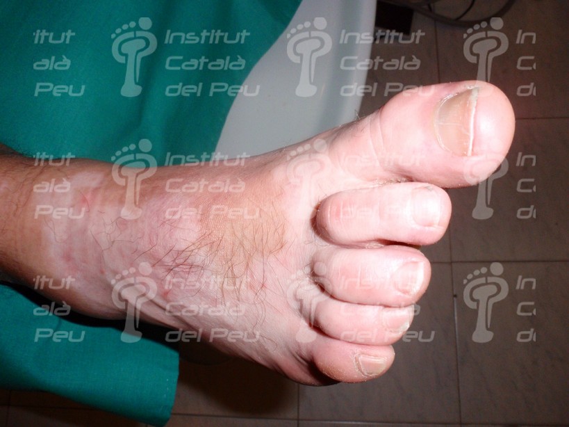
The more frequent spots appear in zones of the skin exposed to the sun (hands, feet, arms, face…) being also common the appearance in the navel, groins and the genital organs. In foot they generally appear in the dorsum and in the malleoli.
The diagnosis of vitiligo is rather clinical. It is diagnosed by wood’s light and sometimes with a biopsy in order to confirm the absence of melanocytes. The wood’s light is an ultraviolet light which provides a bright white color to the hypopigmented cutaneous surface.
The treatment changes according to the number of spots and how much they have spread. The treatments are based in corticosteroids, psoralens together with UVA and immunodepressor topic treatments, having them a slender result. It is recommended strong sunblocks because the absence of pigmentation increases the risk of lesions by solar radiation. In some specific cases the grafts through surgery may be assessed.


Trackbacks & Pingbacks
… [Trackback]
[…] Find More Informations here: institutcataladelpeu.com/en/the-vitiligo-definition-diagnosis-and-treatment-of-this-achromic-lesion/ […]
Leave a Reply
Want to join the discussion?Feel free to contribute!