BIPARTITE PROXIMAL PHALANX
The primary center of ossification is made through deposits of a transverse disk in the cartilage matrix, approx. at the level of the embryonal diaphysis center. The center of this calcified zone is immediately absorbed and it is then changed in the marrow canal. The width of the marrow canal increases at the same time than the large bore of diaphysis does. The secondary center usually appears after birth. The cartilaginous stripe interposed between diaphysis and the center of ossification, progressively decreases its thickness until it completely disappears when growth finishes. At this moment the epiphysis and the diaphysis join together in order to make up the adult bone. The normal ossification of the epiphyseal center is not an uniform process, because instead of an only center it can appear several osseous focus of small size which they later join together and make up a bigger osseous contour and of a bigger size. All the bones of the foot are being formed from a primary center, while phalanges, metatarsal and calcaneal have moreover, an ossification secondary center. The secondary nucleus of ossification shows a partial metaphyseal closing state at 12 years old until the possibility of 16 years old. In the fetal period ossification begins in the metatarsals followed by the distal, proximal and finally, the medium phalanx.
According to the stood clinical situation, we have some ossification trouble in the proximal phalanx of the first toe, thus I am more interested in observing the ossification of phalanges.
Phalanges begin to be seen by radiology in a proximal-distal and medium-lateral direction. The distal phalanx of first toe begins to be observed in the ninth week of the fetal life and continues through the distal phalanges of the remaining toes during the eleventh and twelfth week and through the proximal phalanges in the fourteenth week. It finishes through the medium phalanges at the end of the fourth month of the uterine life.
According to the provided survey it is considered that:
- The first secondary nucleus of ossification which appears is from the distal phalanx of first toe, that is to say, at two and two month years old and a bit later it is formed the secondary nucleus of distal phalanges of the remaining toes.
- In the first radius, the proximal phalanx receives the vascular contribution from the dorsal digital artery while the distal phalanx irrigates through the plantar artery.
I put a special emphasis in the growth of the foot’s size, where I can see that in birth, the foot’s dimension is between 20 and 34 % of the final adult size, being its size approx. 7 and 8 cm. In baby girls, about 12 months old and baby boys about 18 months old, foot raises half of the final length and at two years old the longitudinal arc of foot is completely developed (Tachdjian 1972 and 1999, Scheuer & Black 2000). This children’s foot between 1 a d 5 years old grows 0,9 cm. per year. At six years old foot has a double length (Vilato 1969). Between 5 and 10 years old foot catches up with 63 % of the whole growth and at 10 years old foot has about 81 % of the final length. At 14 years old, girls catch up with such length and boys at 15 years old, although Dr. Tachdjian states this theory in girls is at 12 years old and in boys at 14 years old and Dr. Robles asserts that in girls is at 13 years old and in boys at 15 years old (Robles 1997).
This means that in all periods of growth the size of foot is nearly the same than in adult size. Thus, factors which may change the growth of foot will affect very few the final size of foot, fact which doesn’t happen in the femur or the tibia (Tachdjian 1972. Ogden (2) 2000). The details in which most of the publications are based on come from the research made by Blais, Green and Anderson in 1956, who established the normal standards of the foot’s growth length after making a survey in 512 children between 1 and 18 years old, among American people in the 50’s (Blais et Al 1956), but as you can see, the ages in which it is produced generates constant controversies.
The bipartite proximal phalanx in an adolescent, sometimes is similar to a fracture, where you can think it may be caused by a lack of attachment in both bones, which can lead to a wrong diagnosis or even to splint unnecessarily the toe, or being operated surgically, bringing lesions in the growth cartilage with the total halt of the bone. Thus, we must consider the diagnosis very much and make radiographies in both feet to be able to compare and not to get wrong with a traumatism or Thiemann’s disease. In a nutshell, the best to distinguish is to watch carefully the radiography, the margines of the bone and in case it is clear, then it will be probably a bipartite bone.
There are few cases of wedges and bipartite phalanges, but according to the surveys which exist, we can say that those cases regarding to phalanges and wedges are more common in men and they are usually bilateral, strongly indicating the genetic component.
In a few words, after all the found and consulted bibliography I can see there are disagreements in the ossification chronology of the wedges (finally, appearing the third one later), primary and secondary ossification nuclei, chronology in the foot’s bones growth and moreover, great contraindications regarding the appearance of the distal phalanx of the fifth radius.
The congenital malformation of first proximal phalanx is because of a lack of fusion or ossification of the primary and secondary nuclei of the phalanx, which this lack of union entails to produce the bipartite.
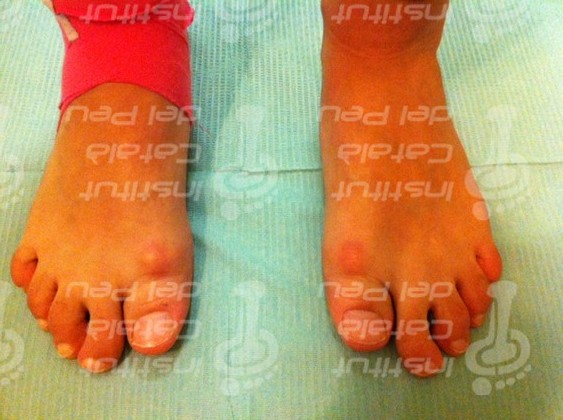
The orthopedic treatment is effective for this kind of pathologies and must lead to reduce the patient’s symptomatology. The pressure of the proximal phalanx of the first radius in both feet has diminished.
If the treatment didn’t work in several years or the patient wouldn’t notice any relief with the orthopedic treatment, then it could be optional a surgical operation. In this case, the most valid and recommended option by the orthopedic surgeons, would be to join the two sides of the phalanx through a compression screw in order to give more stability to this phalanx, but obviously, the best is to wait that our adolescent patient has finished the whole growth and so reevaluate the whole situation according to the sequelae which he shows.

Radiological view of the proximal phalanx of the first toe, bipartite and bilateral.

Dorsum-plantar radiography of the right foot. Watch that the bipartite phalanx is smaller.

Supporting surface above the podoscope. Watch the disability of the first radius.

Anteroposterior view in charge.

Anteroposterior view in the podoscope. Watch the charge transfer in the second radius.


Trackbacks & Pingbacks
Milwaukee Bucks Jerseys UK
Data for all inspected balls, including the amount of air in each, will be sent to the league office for evaluation.
Leave a Reply
Want to join the discussion?Feel free to contribute!