The subungual heloma is a commonly keratosis known as callosity which is located in the nail bed and underlying to the unguinal layer. It appears statistically most of the time on the first toe.
The causes which help the appearance of the subungual heloma are several:
Repetitive politraumatisms by the use of shoes with short toe.
Hallux extensus by the traction of the common extensor of the first toe, which falls within the basis of the proximal phalanx.
Appearance of exostosis or subungual osteophyte. (It is diagnosed by a profile radiologic image).
Egyptian foot.
Certain physical activities.
Some of the features of the subungual heloma are the appearance of a circumscribed zone of red or light brown subungual color and a probable thicknening of the unguinal layer by hypertension and microtraumatisms.
It is necessary to do a differential diagnosis against a hypothetical chondroma or subungual exostosis. Sometimes, the patient has the wrong sensation of having an ingrown toenail, because when putting pressure on the unguinal layer, causes pain of difficult location.
Watching frontally the unguinal layer, you can notice the appearance of a fine hyperkeratosis between the nail bed and the corresponding layer to the unguinal heloma.
Initially, it is carried out a conservative treatment, which lies in the exeresis of the unguinal portion which covers the heloma and the enucleation of the latter by scalpel. In those cases of hyperextension of the toe, it can be assessed a silicone orthesis in order to prevent such hyperextension.
The surgical treatment is another option depending on the frequency and intensity of pain which the subungual heloma will cause.
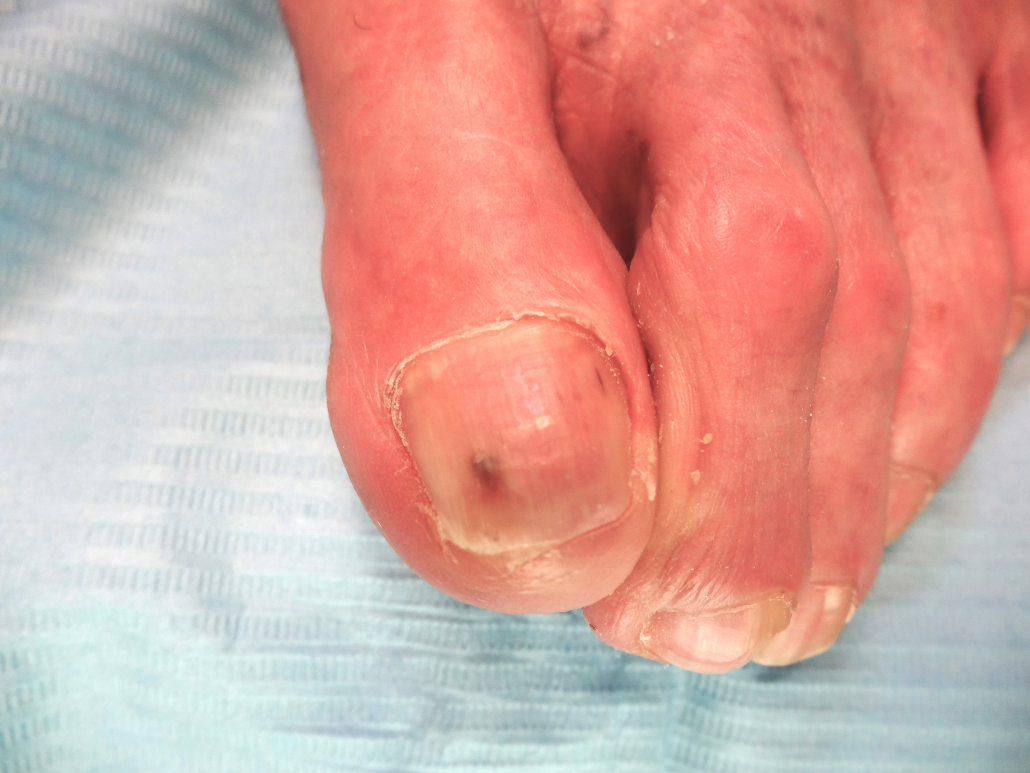
Subungual heloma. Watch the appearance of a small hematoma next to the heloma.
Enucleation of the subungual heloma of the same previous case through exeresis of the unguinal layer in wedge.
Image of a heloma after removing the unguinal layer in wedge.
Visualization of subungual heloma after removing the unguinal layer caused by the second supraductus toe.
Profile x-ray where you can see exostosis in dorsum of the distal phalanx.
Subungual heloma. Sometimes it can be confused with an ingrown toenail.
Visualization of a subungual heloma after removing the previous part of the unguinal layer.
Enucleation of the subungual heloma of the previous case.
First toe once the subungual heloma is enucleated.
0replies
Leave a Reply
Want to join the discussion? Feel free to contribute!
INSTITUT CATALA PODOLOGIA, SL utiliza "COOKIES" para garantizar el correcto funcionamiento de nuestro portal web, mejorando la seguridad, para obtener una eficacia y una personalización superiores, para recoger datos estadísticos y para mostrarle publicidad relevante. Si continúa navegando o pulsa el botón "ACEPTAR" consideraremos que acepta todo su uso. Puede obtener más información en nuestra POLÍTICA DE COOKIES en el pie de página.


Leave a Reply
Want to join the discussion?Feel free to contribute!