THE NEUROPATHIC ARTHROPATHY OR CHARCOT’S FOOT.
The neuropathic arthropathy is also known as Charcot’s foot as an acknowledgment to Jean-Marie Charcot, the first man who described this chronic and degenerative neuropathy. The neuropathic arthropathy is the result of different peripheral neuropathies, being the diabetic neuropathy the main cause.
The main features of the Charcot’s foot are the appearance of an erythema or reddening of the skin, localized intense inflammation with osseous debility which bears sprains, as well as osseous destruction, subluxation and deformity of the foot or even on the ankle. All this, is a consequence of several factors, like for example, the diabetes, the motor- sensitive neuropathy, the autonomic neuropathy and osseous metabolic disorders.
The Charcot’s foot has an inflammation or widespread edema of foot next to a neural vascular reflex which implies a permanent vasodilatation with the consequent active resorption of the osseous tissue because of loss of the sympathetic innervation.
This constant vasodilatation causes a prime feature of Charcot’s foot, which is an increase of the cutaneous temperature.
Although on acute stages, the patient who suffers neuropathic arthropathy may have pain, in most of the cases it usually falls. The peripheral sensorial neuropathy with the fall of pain are crucial in Charcot’s foot.
The osseous remodeling with the appearance of osseous prominences by the lack of feeling is a very high risk of formation of lesions in the foot or ankle and as a result, superficial or deep infections of these lesions, possibly becoming to amputations. The most frequent osseous deformities are digital, splay foot and even osseous remodeling of the foot with a rocking chair shape on late stages.
The neuropathy is a common feature of Charcot’s foot, being diabetes a crucial cause of the neuropathy.
The 80 % of diabetic patients who suffer neuropathic artropathy are diagnosed of diabetes for more than ten years and most of them don’t have a good control.
Other factors which help this neuropathy are alcoholism, traumatisms, the syringomyelia, Parkinson, VIH, Hansen’s disease or rheumatic diseases.
The patient usually notices structural changes in his foot, even crepitations of the bones when wandering around.
According to Eichenholtz, the neuropathic arthropathy is classified in 3 stages:
- First stage. Development of the arthropathy. Osseous destruction and fragmentation with hyperemia and cutaneous trophic changes. The foot is tumescent and hot.
- Second stage. Coalescence. Beginning of the osseous repair as well as progressive disappearance of some symptoms (rubor, edema and heat).
- Third stage. Osseous consolidation with residual deformity.
An X-ray can be used in order to get an accurate diagnosis, with the aim of visualizing fractures or subluxations, calcification of arteries and reduction of the calcaneal inclination angle on advanced stages. The changes of the Charcot’s foot in a radiological level are belated, therefore, a magnetic resonance should be recommended in order to detect minimum changes on early stages. It is important not to forget the medical history, the physical exploration and the analysis laboratory.
The purpose of the conservative treatment is to reduce the foot’s edema and to prevent possible fractures. The edema and the rise in temperature are reduced through the immobilization with complete plaster at least for 18 weeks, which must be periodically replaced until the temperature of both feet is equal. Once this stage is finished, orthesis, discharge splints and suitable shoes are used during the wandering about depending on the different levels of affectation.
The surgical treatment must be made to correct deformities, mainly the joint of the ankle, disarticulation or osteomyelitis.
On developed stages of the Charcot’s foot, surgery must not be used. The aim of surgery is the use of bone grafts and arthrodesis with the purpose of stabilizing and restoring the position and functions, foreseeing subsequent osseous destructions.

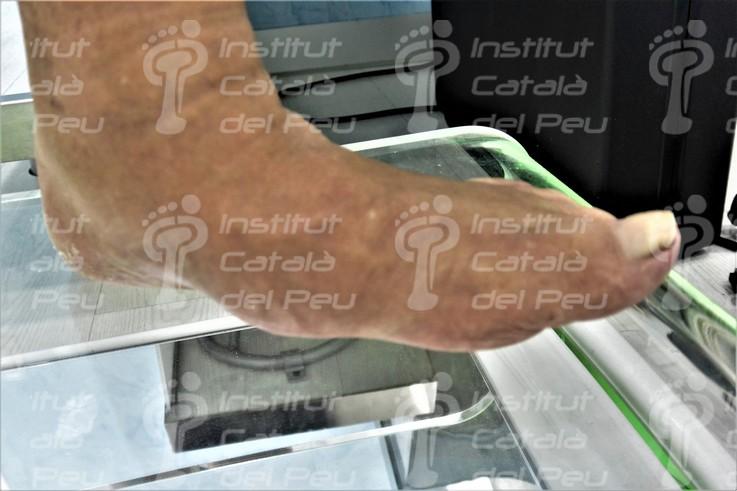
Charcot’s foot. You can watch the osseous pronation of the plantar external arch.

The Charcot’s foot causes serious changes of the plantar support.

Profile X-ray of Charcot’s foot. You can watch the image in a rocking chair shape.

Dorsum plantar X-ray. You can watch the affectation on left foot.

Complication of Charcot’s foot. Plantar ulcer.

Complication of Charcot’s foot. Plantar ulcer with osteomyelitis.

Affectation of fifth metatarsal in Charcot’s foot. You can watch the osseous remodeling which the fourth toe suffered.

Complication of Charcot’s foot. Plantar ulcer.

Discharge plantar support.

Walker’s boot.


Leave a Reply
Want to join the discussion?Feel free to contribute!