


Surgery is recommended in those cases in which the osteoma causes discomfort and also when it causes aesthetic deformities as is usually the case in subungual osteomas (they are quite often young patients). The most important thing in this kind of surgery is to curette the osseous surface once the osteoma has been excised, in order to avoid a recidivation risk. It is imperative to make this surgery with X-ray support, to make sure that the proper osseous scaling has been carried out before finishing theoperation and obviously sending the excised tumors to pathologic anatomy in order to confirm the diagnosis completely.
From one of my cases, I will describe you schematically the steps to follow in this kind of surgery.

aspect of osteoma in the fourth toe.

toe nerve block anesthesia.

osteoma delimitation.

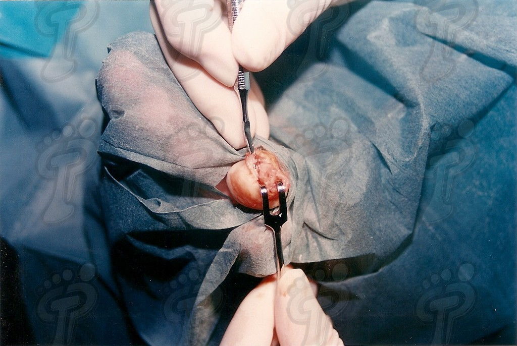
cut of tumor by metal shears.

cut of tumor by metal shears.

curettage with Jansen small spoon.

scaling of osseous cortical by a lime.

scaling of osseous cortical by a lime.

cleaning by dragging with physiologic serum.

surgical wound in 48 hours.

postoperative stage in six days.

medical discharge in hospital in eleven days.